Assay Protocol
Protocol
For 200, 500, and 2500 reactions
Store at -20°C upon arrival

1. General Information
The performance of the EpiMelt Methylation Detection Assay has been tested on the LightCycler® 480
System (Roche, catalog no. 05015278001) with the EpiMelt Real-Time PCR Master Mix (MethylDetect, catalog no EPI-pPCR-200) in 96-well plates.
The use of the EpiMelt Methylation Detection Assay in combination with this platform is recommended.
! Other RT-PCR systems capable of performing high-resolution melting (HRM)
can be used, provided the experimental conditions are optimized before sample testing !
For details on RT-PCR setup and optimization of experimental conditions, please refer to the Preparation of the PCR reaction paragraph in section 2. Protocol.
Contents
| Label | Amount per assay | Amount per vial (200 reactions) | Amount per vial (500 reactions)* |
| EpiMelt Primer Mix | 1 vial | 240 μl | 550 μl |
| EpiMelt Methylation Positive Control | 1 vial | 70 μl | 125 μl |
| EpiMelt Assay Calibration Control | 1 vial | 70 μl | 125 μl |
| EpiMelt Methylation Negative Control | 1 vial | 70 μl | 125 μl |
Table 1. Contents of the EpiMelt Methylation Detection Assay.
*: For 2500 reactions: 5 x EpiMelt Methylation Detection Assay (500 reactions) is sent.
Storage conditions
The EpiMelt Methylation Detection Assay is shipped at room temperature. When stored at – 20°C, the reagents are stable through the expiration date. Shipping and temporary storage at 4°C after opening for up to 8 weeks have no detrimental effects on the assay performance.
Avoid repetitive freezing and thawing of the assay components.
For storage conditions of the EpiMelt Real-Time PCR Master Mix, see the associated protocol.
Additional equipment and reagents required:
- EpiMelt Real-Time PCR Master Mix
- EpiMelt Master Mix (Containing a hot-start DNA polymerase, reaction buffer, dNTP mix, HRM dye, and MgCl2)
- PCR-grade H2O
- A Real-Time PCR instrument capable of HRM
- Pipettes with sterile, nuclease-free, aerosol-resistant filter tips
- Sterile reaction tubes for preparation of PCR mix
- Standard swinging-bucket centrifuge for multi-well plates (optional)
Application
The EpiMelt Methylation Detection Assay is designed and optimized for the detection of the DNA methylation status of a specific gene promoter region in human bisulfite-converted genomic DNA. Three controls are supplied with the assay, containing a methylation positive control, a methylation negative control, and an assay calibration control, to ensure easy data analysis and optimal performance of the assay.
Assay time
The cycling program for the EpiMelt Methylation Detection Assay includes 10 min pre-incubation, followed by 50 amplification cycles and HRM. In total, this is approx. 90-120 min when performed on the LightCycler® 480 System.
2. Protocol
Before you begin
Sample materials
When using PCR for methylation studies, all DNA samples have to be bisulfite converted to ensure the preservation of the methylated cytosines in the template. The changes induced in the DNA by the sodium bisulfite treatment are illustrated in Figure 1:
Template DNA:

Bisulfite converted DNA:

Figure 1. The underlined and red bases represent CpG dinucleotides containing a methylated cytosine, which is preserved during bisulfite treatment. The blue bases represent unmethylated cytosines, which are converted to uracil during bisulfite treatment and subsequently replaced by thymine during PCR.
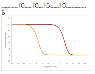
Bisulfite treatment converts unmethylated cytosines into uracil and leaves the methylated cytosines unchanged. The DNA sequence of the methylated DNA strand is therefore different from the unmethylated strand. This results in different melting properties of the two PCR products, which are clearly distinguishable after HRM (Figure 2).
A
Un-methylated sequence
A
Methylated sequence
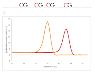
Figure 2. Schematic illustration of the principle behind HRM analysis. A) The difference in a DNA sequence after bisulfite conversion of a methylated and an unmethylated region. B) The difference in melting properties of the PCR products from the methylated (red) and the unmethylated (orange) templates displayed as melting curves and melting peaks.
Sample DNA
Use 30-50 ng of bisulfite-modified DNA per PCR reaction. This is a theoretical concentration based on the DNA amount used for bisulfite conversion and the elution volume. The EpiMelt Bisulfite Modification Standard Kit is recommended for the bisulfite conversion of sample DNA.
! Follow the instructions from the bisulfite conversion kit for details on DNA storage after bisulfite conversion and clean-up !
The quality of the DNA should be suitable for PCR in terms of concentration, purity, and absence of PCR inhibitors. Use the same DNA extraction procedure for all samples in the analysis. To ensure sufficient quality DNA prior to bisulfite conversion, analysis of DNA integrity and concentration is recommended.
Assay controls
A methylation positive control, a methylation negative control, and an assay calibration control are supplied with the EpiMelt Methylation Detection Assay. The assay calibration control is included to ensure appropriate assay sensitivity.
! The supplied controls are ready to use and should not be bisulfite converted !
No-template control
A No-template control (NTC) should always be included in the analysis. The NTC contains the same reagents as the reactions for analysis, except that the DNA sample is replaced by an equal amount of PCR-grade water. The NTC should be included in triplicates in each multi-well plate.
Primers
A primer mix is supplied with the EpiMelt Methylation Detection Assay. The PCR assay conditions are listed in Table 2.
Preparation of the PCR reaction
This protocol is optimized using the EpiMelt Real-Time PCR Master Mix on the LightCycler® 480 System. If used on an alternative system, the annealing temperature should be optimized prior to sample testing. This can be done using the controls supplied with the EpiMelt Methylation Detection Assay. Follow the procedure below in the given order to prepare one 20 μL standard reaction.
- Thaw the solutions on ice, vortex, and spin briefly, to ensure that the content is collected at the bottom of the tube.
- Prepare the PCR mix for one 20 μL reaction by adding the following components in the order listed in Table 1.
| Component | Volume |
| EpiMelt Real-Time PCR Master Mix 2x | 10 μl |
| EpiMelt Primer Mix | 1 μl |
| EpiMelt PCR-grade H2O | 3 μl |
| Total | 14 μl |
Table 2. Components of the PCR mix
Multiple reactions
To prepare the PCR mix for multiple reactions, multiply the volume of each component by the total number of reactions. The total number of reactions should include samples to be analyzed, controls supplied with the assay, and the NTC.
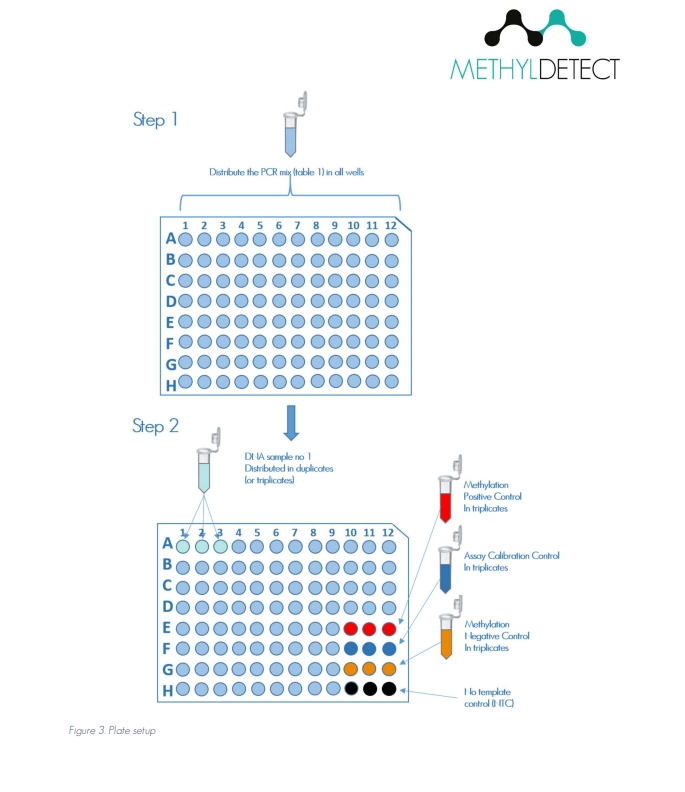
! For an example of plate setup, see Figure 3 !
Example of PCR plate preparation
Preparation of a 96-well plate, in which all samples are analyzed in triplicates:
- 28 test samples in triplicates → 84 reactions. Three assay controls in triplicates → 9 reactions. NTC in triplicates → 3 reactions. In total: 96 reactions.
- Calculate the volume needed from each reagent and add them to a tube to make the PCR mix. Mix by pipetting up and down. Do not vortex.
- Pipette 14 μL PCR mix into each well in a 96-well plate.
- Add 6 μL bisulfite converted sample DNA to the appropriate wells, corresponding to a calculated volume of 30-50 ng DNA. It is possible to use a lower amount of template. However, we recommend that the assay is optimized to the specific DNA concentration before processing the test samples.
- Add 6 μL of each standard to the appropriate wells.
- Seal the multi-well plate with an appropriate sealing foil.
- Spin for 2 min at 1000xg.
- Place the multi-well plate in the LightCycler® 480® instrument and start the PCR-HRM program.
Download the Protocol here:
| Program | Cycles | Temperature (°C) | Acquisition mode | Hold (sec) | Ramp rate (°C/sec) | Aquisitions (per °C) |
| Pre-incubation | 1 | 95 | None | 600 | 4.4 | – |
| Amplification | 50 | 95 * 72 |
None None Single |
15 10 15 |
4.4 2.2 4.4 |
– – – |
| High-Resolution Melting | – | 95 60 95 |
None None Continuous |
15 60 – |
4.4 2.2 0.01 |
– – 50 |
Table 3. PCR and HRM program. *: Temperature is assay-specific and may differ between platforms. The program is suitable for the LightCycler® 480 System.
3. The principle behind the MS-HRM analysis method
Methylation-Sensitive High-Resolution Melting (MS-HRM) is a high-throughput technology for highly sensitive, closed-tube analysis of locus-specific methylation1,2. The technology utilizes the difference in melting properties of the PCR product amplified from methylated and unmethylated DNA strands after bisulfite conversion (Figure 1).
The proprietary design of the EpiMelt Methylation Detection Assays makes them suitable to differentiate between methylated, unmethylated, and mixed templates, and to distinguish heterogeneously methylated templates3-5.
4. Results
The results presented below were obtained using an EpiMelt Methylation Detection Assay and the EpiMelt Real-Time PCR Master Mix. After amplification, the PCR product is subjected to HRM analysis, and the data is analyzed using the LightCycler® 480 Gene Scanning or Tm-Calling Software.
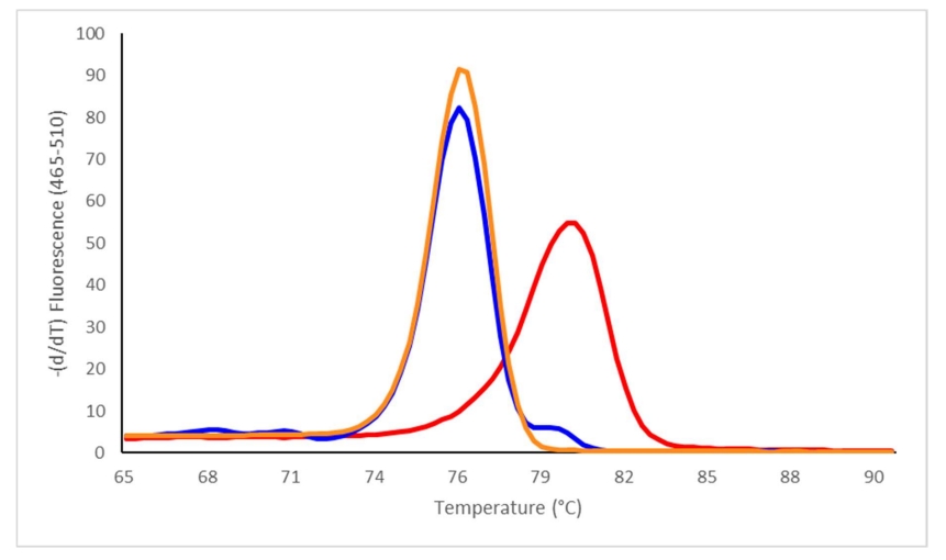
Figure 4. Relative signal difference (-d/dT) plot illustrating the melting profiles of the methylation positive control (red), the methylation negative control (orange), and the assay calibration control (blue) supplied with the EpiMelt Methylation Detection Assay.
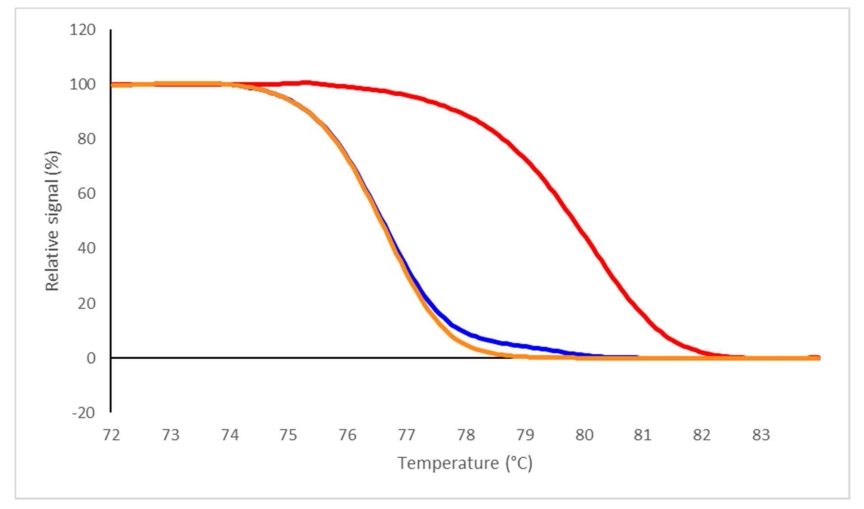
Figure 5. Relative signal difference (-d/dT) plot illustrating the melting profiles of the methylation positive control (red), the methylation negative control (orange), and the assay calibration control (blue) supplied with the EpiMelt Methylation Detection Assay.
Download the Protocol here:
5. References
- Clark, S. J., Harrison, J., Paul, C. L. & Frommer, M. High sensitivity mapping of methylated cytosines. Nucleic Acids Res 22, 2990-2997 (1994). https://doi.org:10.1093/nar/22.15.2990.
- Wojdacz, T. K., Dobrovic, A. & Hansen, L. L. Methylation-sensitive high-resolution melting. Nature
protocols 3, 1903-1908 (2008). https://doi.org:10.1038/nprot.2008.191. - Wojdacz, T. K. & Dobrovic, A. Methylation-sensitive high resolution melting (MS-HRM): a new approach for sensitive and high-throughput assessment of methylation. Nucleic Acids Res 35, e41 (2007). https://doi.org:10.1093/nar/gkm013.
- Wojdacz, T. K. et al. Abstract 2308: Heterogeneous and low-level methylation of novel biomarker candidates for breast cancer clinical management. Cancer Research 74, 2308-2308 (2014). https://doi.org:10.1158/1538-7445.Am2014-2308.
- Hussmann, D. & Hansen, L. L. Methylation-Sensitive High Resolution Melting (MS-HRM). Methods in molecular biology (Clifton, N.J.) 1708, 551-571 (2018). https://doi.org:10.1007/978-1-4939-7481-8_28.
6. Troubleshooting
| Observation | Possible cause | Recommendation |
| No detectable amplifications | High-Resolution Melting Master was not sufficiently activated. | Make sure that the PCR protocol contains an initial pre-incubation step of 10 min at 95°C |
| No detectable amplifications | Wrong filter combination was used to display the amplification | Select the appropriate filter combination for your EpiMelt Assay on the analysis screen and start again (LightCycler®480 System only) |
| No detectable amplifications | Wrong detection format was chosen for the experimental protocol | Select the appropriate detection format for your EpiMelt Assay and start again |
| No detectable amplifications | Pipetting errors or omitted reagents | Check for missing or impaired reagents |
| No detectable amplifications | Some reagents are still in the upper part of the microwell, or an air bubble is trapped in the reagents | Check the multi-well plate before PCR. Repeat the centrifugation for 2 min at 1000xg if necessary. |
| Fluorescence intensity variable or too low | Deterioration of the dye as the HRM master mix was not stored properly | Store the HRM master mix at -20°C and keep away from light. Avoid freeze-thaw cycles. |
| Negative control sample gives an increasing signal during PCR | Reagents may be contaminated | Use contamination-free components |
| Negative control sample gives an increasing signal during PCR | Reagents may be contaminated | Use contamination-free components |
| Curve shapes in the Relative signal difference plot are variable without recognizable pattern; no reasonable grouping | Concentration or quality of the DNA is not consistent |
The lowest signal increase during amplification must not be less than 60% of the highest. The signal levels at the beginning of the melting curve should be as similar as possible. _________________________________________________ Assure that amplification curves show Cq values <30 cycles and are in a range of less than 3 cycles for all samples. |
7. Ordering information
MethylDetect offers a number of EpiMelt Methylation Detection Assays targeting specific genomic regions. For a complete overview of the products and ordering information, please visit www.MethylDetect.com.
8. Supplementary information
License disclaimer
For patent license limitations for individual products, please refer to www.MethylDetect.com.
Regulatory disclaimer
For Life Science research only. Not for use in diagnostic procedures.
Safety data sheet
Please follow the instructions in the safety data sheet (SDS) at www.MethylDetect.com.
Contact and support
Please refer to www.MethylDetect.com.
! For research use only. Not for use in diagnostic procedures !
User manual version 3. Last revision date: 23/06/2023
©2019 MethylDetect. All rights reserved.
